- 0086-571-85302990
- sales@greenskybio.com


Enhancing Plant Research with Actin: A Detailed Protocol for Protein Extraction and Western Blot Analysis
2024-08-02
1. Introduction
Actin is one of the most important cytoskeletal proteins in plants. It plays a crucial role in a wide range of plant physiological processes. These include cell division, cell elongation, cytoplasmic streaming, and organelle movement. Understanding the function and regulation of actin at the molecular level is essential for advancing plant research. Protein extraction and Western blot analysis are powerful techniques that can be used to study actin in plants. In this article, we present a detailed protocol for these techniques, which will be valuable for researchers in the field of plant biology.
2. Significance of Actin in Plant Physiological Studies
2.1 Cell Division
During cell division in plants, actin filaments form a contractile ring that is involved in cytokinesis. This process is essential for the proper partitioning of the cytoplasm and the formation of two daughter cells. Actin also plays a role in spindle formation and chromosome movement during mitosis. Any disruption in actin function can lead to abnormal cell division and ultimately affect plant growth and development.
2.2 Cell Elongation
Cell elongation is a key process in plant growth, especially in the growth of roots and shoots. Actin filaments are involved in the orientation and expansion of cells. They help to maintain the shape of the cell and also play a role in the transport of vesicles and cell wall components to the site of cell elongation. Mutations in actin - related genes can result in stunted growth or abnormal cell shapes.
2.3 Cytoplasmic Streaming
Cytoplasmic streaming is the movement of the cytoplasm within a cell. In plants, this process is driven by actin - myosin interactions. Cytoplasmic streaming is important for the distribution of nutrients, organelles, and signaling molecules within the cell. It also plays a role in the response of plants to environmental stimuli. For example, in response to light, cytoplasmic streaming can be altered to optimize photosynthesis.
2.4 Organelle Movement
Actin filaments are involved in the movement of organelles such as mitochondria, chloroplasts, and peroxisomes within plant cells. This movement is important for the proper functioning of these organelles. For example, the movement of chloroplasts in response to light intensity helps to optimize photosynthesis. Actin - related proteins also play a role in the targeting of organelles to specific locations within the cell.
3. Protein Extraction Protocol
3.1 Materials
- Plant tissue (fresh or frozen)
- Extraction buffer (e.g., Tris - HCl buffer, pH 7.5, containing protease inhibitors)
- Mortar and pestle (for grinding plant tissue)
- Centrifuge tubes
- Centrifuge
3.2 Procedure
- Harvest plant tissue: Select the appropriate plant tissue for analysis. For example, if studying root growth, harvest the root tips. Ensure that the tissue is fresh or properly frozen to preserve protein integrity.
- Grind the tissue: Place the plant tissue in a mortar and add a sufficient amount of extraction buffer. Use a pestle to grind the tissue into a fine paste. This step helps to break open the cells and release the proteins.
- Transfer the homogenate: Transfer the ground tissue homogenate to a centrifuge tube. Make sure to transfer all of the homogenate to ensure accurate protein extraction.
- Centrifuge the homogenate: Centrifuge the homogenate at a high speed (e.g., 10,000 - 15,000 x g) for 10 - 15 minutes. This step separates the supernatant (which contains the soluble proteins) from the pellet (which contains cell debris and insoluble proteins).
- Collect the supernatant: Carefully transfer the supernatant to a new centrifuge tube. The supernatant is the protein extract that will be used for further analysis.
4. Western Blot Analysis Protocol
4.1 Materials
- Protein extract from previous step
- SDS - PAGE gel (sodium dodecyl sulfate - polyacrylamide gel electrophoresis)
- Transfer buffer
- Nitrocellulose or PVDF (polyvinylidene fluoride) membrane
- Blocking buffer (e.g., 5% non - fat dry milk in Tris - buffered saline - Tween 20, TBST)
- Primary antibody against actin
- Secondary antibody (conjugated to a detectable label, e.g., horseradish peroxidase)
- Chemiluminescent substrate
- X - ray film or a digital imaging system
4.2 Procedure
- Prepare the SDS - PAGE gel: Cast the SDS - PAGE gel according to the manufacturer's instructions. This gel will be used to separate the proteins in the extract based on their molecular weights.
- Load the protein samples: Load an appropriate amount of the protein extract onto the SDS - PAGE gel. Also, load a molecular weight marker for accurate protein size determination.
- Run the gel: Run the SDS - PAGE gel at a constant voltage (e.g., 100 - 150 V) until the dye front reaches the bottom of the gel. This process separates the proteins in the extract into distinct bands based on their molecular weights.
- Transfer the proteins: Transfer the separated proteins from the SDS - PAGE gel to a nitrocellulose or PVDF membrane using a transfer apparatus and transfer buffer. This step is crucial for immobilizing the proteins on the membrane for subsequent antibody binding.
- Block the membrane: Incubate the membrane in blocking buffer for 1 - 2 hours at room temperature or overnight at 4°C. Blocking helps to prevent non - specific binding of antibodies to the membrane.
- Incubate with primary antibody: Incubate the membrane with the primary antibody against actin in a suitable dilution (e.g., 1:1000 - 1:5000) in blocking buffer for 1 - 2 hours at room temperature or overnight at 4°C. The primary antibody will specifically bind to actin proteins on the membrane.
- Wash the membrane: Wash the membrane several times (e.g., 3 - 5 times) with TBST to remove unbound primary antibody.
- Incubate with secondary antibody: Incubate the membrane with the secondary antibody in a suitable dilution (e.g., 1:5000 - 1:10000) in blocking buffer for 1 - 2 hours at room temperature. The secondary antibody will bind to the primary antibody, creating a detectable complex.
- Wash the membrane again: Wash the membrane several times with TBST to remove unbound secondary antibody.
- Detect the signal: Incubate the membrane with a chemiluminescent substrate. The substrate will react with the enzyme conjugated to the secondary antibody, producing a chemiluminescent signal. This signal can be detected using X - ray film or a digital imaging system. The presence and intensity of the signal indicate the amount of actin protein in the sample.
5. Conclusion
Actin is a fundamental component of plant cells, and its study is of great importance in plant research. The protein extraction and Western blot analysis protocols described in this article provide a reliable and efficient way to study actin at the molecular level. By following these protocols, researchers can gain insights into the function and regulation of actin in various plant physiological processes. This knowledge can be further applied to improve plant growth, development, and stress tolerance, ultimately contributing to the advancement of plant biology research.
FAQ:
1. Why is actin important in plant research?
Actin plays a crucial role in various aspects of plant physiology. It is involved in cell structure and shape maintenance, as it is a major component of the cytoskeleton. Actin also participates in cell division, cell expansion, and intracellular transport processes. In plant research, studying actin helps in understanding these fundamental processes at the molecular level, which is essential for overall understanding of plant growth, development, and responses to environmental stimuli.
2. What are the key steps in the protein extraction protocol for actin?
The key steps in the protein extraction protocol for actin typically include sample collection, which should be done carefully to ensure the relevant plant tissues are obtained. Then, the tissues are usually homogenized in an appropriate buffer. This buffer is designed to break open the cells and keep the proteins stable. After homogenization, centrifugation is carried out to separate the supernatant (containing the proteins) from the cell debris. The supernatant is then collected for further analysis. Different plant species or tissues may require some adjustments in the buffer composition and extraction conditions to optimize the extraction of actin - related proteins.
3. How can one ensure accurate Western blot analysis for actin?
To ensure accurate Western blot analysis for actin, several aspects need to be considered. First, the quality of the protein samples is crucial. This means proper extraction and storage to prevent protein degradation. Second, the choice of antibodies specific to actin is very important. High - quality and specific antibodies will increase the accuracy of detection. During the electrophoresis step, appropriate running conditions should be set to separate the actin proteins clearly. Also, proper transfer conditions to the membrane are necessary to ensure efficient transfer of the proteins. Finally, the detection method, such as the use of chemiluminescent substrates, should be optimized to get clear and reliable signals.
4. Are there any challenges in the protein extraction and Western blot analysis of actin in plants?
Yes, there are several challenges. One challenge is the presence of interfering substances in plant tissues. These substances can affect the extraction efficiency and the accuracy of Western blot analysis. For example, some plant metabolites may bind to the proteins or interfere with the antibodies. Another challenge is the variability in actin protein levels among different plant tissues and developmental stages. This requires careful selection of samples and normalization methods. Additionally, the complexity of the plant cytoskeleton, which contains multiple isoforms of actin, can make it difficult to specifically detect and analyze a particular form of actin.
5. How can the results of actin protein extraction and Western blot analysis be applied in plant research?
The results of actin protein extraction and Western blot analysis can be applied in multiple ways in plant research. Firstly, it can help in understanding the regulation of actin expression during different developmental stages of plants, such as during seed germination or flower development. Secondly, by studying changes in actin levels in response to environmental stresses like drought or salinity, researchers can gain insights into how plants adapt to these adverse conditions at the molecular level. Moreover, comparing actin profiles in different plant genotypes can assist in identifying genetic factors related to plant growth and development.
Related literature
- Title: Actin - Based Motility in Plants: The Role of Actin - Binding Proteins"
- Title: "The Cytoskeleton in Plant Development: Function and Regulation of Actin Filaments"
- Title: "Advances in Actin Research in Plant Physiology: New Insights and Perspectives"


- ▶ Hesperidin
- ▶ Citrus Bioflavonoids
- ▶ Plant Extract
- ▶ lycopene
- ▶ Diosmin
- ▶ Grape seed extract
- ▶ Sea buckthorn Juice Powder
- ▶ Fruit Juice Powder
- ▶ Hops Extract
- ▶ Artichoke Extract
- ▶ Mushroom extract
- ▶ Astaxanthin
- ▶ Green Tea Extract
- ▶ Curcumin
- ▶ Horse Chestnut Extract
- ▶ Other Product
- ▶ Boswellia Serrata Extract
- ▶ Resveratrol
- ▶ Marigold Extract
- ▶ Grape Leaf Extract
- ▶ New Product
- ▶ Aminolevulinic acid
- ▶ Cranberry Extract
- ▶ Red Yeast Rice
- ▶ Red Wine Extract
-
Polygonum Cuspidatum Extract
2024-08-02
-
Saponin Extract
2024-08-02
-
Echinacea Extract
2024-08-02
-
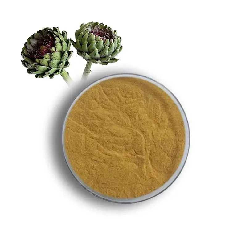
Artichoke Extract
2024-08-02
-
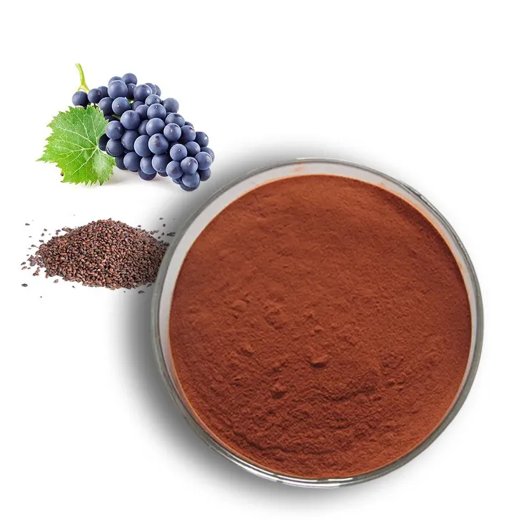
Natural grape seed extract
2024-08-02
-
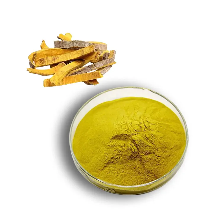
Phellodendron Extract
2024-08-02
-
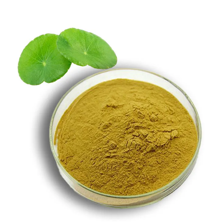
Centella Asiatica Extract
2024-08-02
-
Hawthorn powder
2024-08-02
-
Berberis aristata Extract
2024-08-02
-

Saw Palmetto Extract
2024-08-02
