- 0086-571-85302990
- sales@greenskybio.com


Unveiling the Antimicrobial Efficacy of Plant Extracts: Insights from the Well Diffusion Method
2024-08-16
1. Introduction
In recent years, there has been a growing interest in natural products, particularly plant extracts, for their potential antimicrobial properties. The well - diffusion method has emerged as a valuable tool in assessing the antimicrobial efficacy of these extracts. This method provides a simple yet effective means of determining the ability of plant extracts to inhibit the growth of microorganisms. With the increasing concern over antibiotic resistance and the search for alternative antimicrobial agents, understanding the factors that influence the antimicrobial efficacy of plant extracts becomes crucial.
2. The Well - Diffusion Method
2.1 Principle
The well - diffusion method is based on the principle of diffusion. A sample of the plant extract is placed in a well, which is usually punched into an agar medium that has been inoculated with a specific microorganism. The extract then diffuses into the agar medium. If the extract contains antimicrobial substances, it will inhibit the growth of the microorganism in a zone around the well. The size of this inhibition zone is measured and used as an indicator of the antimicrobial efficacy of the extract.2.2 Procedure
- First, the agar medium is prepared and sterilized. Different types of agar can be used depending on the microorganism to be tested. For example, Mueller - Hinton agar is commonly used for bacteria.
- The agar is then poured into Petri dishes and allowed to solidify.
- The microorganism is inoculated onto the agar surface, either by spreading a suspension of the microorganism evenly or by streaking.
- Wells are punched into the agar using a sterile cork borer or a similar tool.
- The plant extract, which is usually prepared in a suitable solvent such as methanol or ethanol, is then added to the wells. A positive control (such as a known antibiotic) and a negative control (the solvent alone) are also included.
- The Petri dishes are incubated at an appropriate temperature and time for the growth of the microorganism. For most bacteria, incubation at 37°C for 18 - 24 hours is common.
- After incubation, the diameter of the inhibition zones around the wells is measured using a ruler or calipers.
3. Factors Influencing the Antimicrobial Efficacy of Plant Extracts
3.1 Plant Species
Different plant species vary significantly in their antimicrobial properties. For example, plants from the Lamiaceae family, such as mint and basil, are known to possess strong antimicrobial activity. This is due to the presence of various secondary metabolites in these plants. These secondary metabolites can include phenolic compounds, terpenoids, and alkaloids. Phenolic compounds, such as flavonoids and phenolic acids, are often responsible for the antioxidant and antimicrobial properties of plant extracts.- Some plants may produce specific compounds in response to environmental stress or microbial attack, which can enhance their antimicrobial efficacy.
- The genetic makeup of the plant also plays a role in determining the types and amounts of antimicrobial compounds it produces.
3.2 Extraction Methods
The method used to extract the plant material can greatly affect the antimicrobial efficacy of the resulting extract.- Solvent extraction is one of the most common methods. Different solvents have different affinities for various plant compounds. For example, polar solvents like water are good at extracting hydrophilic compounds, while non - polar solvents like hexane are better for lipophilic compounds. Using a combination of solvents can often yield a more comprehensive extract with a wider range of antimicrobial compounds.
- Extraction time and temperature also matter. Longer extraction times and higher temperatures may increase the yield of certain compounds, but they can also lead to the degradation of some sensitive compounds. For example, excessive heat can cause the breakdown of heat - sensitive phenolic compounds, reducing the antimicrobial activity of the extract.
3.3 Types of Microorganisms Targeted
Plant extracts may show different levels of efficacy against different types of microorganisms.- Gram - positive and gram - negative bacteria often respond differently to plant extracts. Gram - positive bacteria have a thicker peptidoglycan layer in their cell walls, while gram - negative bacteria have an outer membrane that can act as a barrier to some antimicrobial compounds. For example, some plant extracts may be more effective against gram - positive bacteria like Staphylococcus aureus, while others may be better at inhibiting gram - negative bacteria such as Escherichia coli.
- Fungi also have unique cell wall structures and metabolic processes compared to bacteria. Some plant extracts are more effective against fungi, such as those containing antifungal compounds like azadirachtin which can disrupt fungal cell membranes or inhibit fungal enzymes.
4. Potential Applications of Plant - Based Antimicrobials
4.1 In the Food Industry
The use of plant - based antimicrobials in the food industry has several advantages.- They can be used as natural preservatives to extend the shelf - life of food products. For example, extracts from plants like rosemary and thyme can inhibit the growth of spoilage bacteria and fungi in food, reducing the need for synthetic preservatives.
- Plant - based antimicrobials can also be used in food packaging. Incorporating antimicrobial plant extracts into packaging materials can help prevent the growth of microorganisms on the surface of the food, improving food safety.
4.2 In the Pharmaceutical Industry
- Plant extracts with antimicrobial properties can serve as a source of new drugs. With the increasing problem of antibiotic resistance, the discovery of new antimicrobial agents from plants is of great importance. For example, some plant - derived compounds are being studied for their potential to treat infections caused by resistant bacteria.
- They can also be used in traditional medicine. Many herbal remedies have been used for centuries to treat various infections, and modern research is now starting to uncover the scientific basis behind their efficacy.
4.3 In Cosmetics
- Antimicrobial plant extracts can be used in cosmetics to prevent the growth of microorganisms in products. This is especially important for products like creams and lotions that are applied to the skin, as they can be a breeding ground for bacteria and fungi if not properly preserved.
- Some plant extracts also have additional beneficial properties for the skin, such as anti - inflammatory and antioxidant effects, making them ideal ingredients for cosmetic products.
5. Conclusion
The well - diffusion method has provided valuable insights into the antimicrobial efficacy of plant extracts. Understanding the factors that influence this efficacy, such as plant species, extraction methods, and the types of microorganisms targeted, is crucial for the development and application of plant - based antimicrobials. With the potential applications in the food, pharmaceutical, and cosmetics industries, plant - based antimicrobials offer a promising alternative to synthetic antimicrobial agents. However, further research is still needed to fully explore the potential of plant extracts, optimize extraction methods, and conduct more in - vivo studies to ensure their safety and efficacy.
FAQ:
What is the well diffusion method?
The well diffusion method is a laboratory technique used to assess the antimicrobial activity of substances, such as plant extracts. In this method, wells are made in an agar medium that has been inoculated with a test microorganism. The plant extract is then placed in these wells. As the extract diffuses into the agar, it inhibits the growth of the microorganism in a circular area around the well. The size of this inhibition zone can be measured and used as an indicator of the antimicrobial efficacy of the plant extract.
How do different plant species affect the antimicrobial efficacy of their extracts?
Different plant species contain a diverse range of bioactive compounds. Some plant species may have a higher concentration of antimicrobial substances compared to others. For example, plants from the Allium genus, like garlic, are known to contain sulfur - containing compounds with strong antimicrobial properties. The chemical composition of plants can vary based on their genetic makeup, which in turn influences the types and amounts of antimicrobial compounds present in their extracts.
What are the common extraction methods for plant extracts?
There are several common extraction methods for plant extracts. One is maceration, where the plant material is soaked in a solvent (such as ethanol or water) for a period of time to allow the extraction of soluble compounds. Another method is Soxhlet extraction, which is a continuous extraction process using a refluxing solvent. Steam distillation is also used, especially for extracting essential oils from plants. The choice of extraction method can affect the yield and quality of the plant extract, and thus its antimicrobial efficacy.
Which types of microorganisms are typically targeted when testing plant extract antimicrobial efficacy?
When testing the antimicrobial efficacy of plant extracts, a wide range of microorganisms are typically targeted. This includes bacteria such as Escherichia coli and Staphylococcus aureus, which are common pathogens in humans. Fungi like Candida albicans are also often studied, as fungal infections can be a significant health problem. Additionally, some research may target plant - pathogenic microorganisms to explore the potential use of plant extracts in agriculture for protecting plants from diseases.
What are the potential applications of plant - based antimicrobials in modern society?
Plant - based antimicrobials have several potential applications in modern society. In the field of medicine, they could be developed into new drugs or used as complementary therapies for treating infections, especially in the face of increasing antibiotic resistance. In the food industry, they can be used as natural preservatives to extend the shelf - life of food products. In cosmetics, plant - based antimicrobials can be added to products to prevent microbial growth. They also have potential applications in agriculture for protecting plants from microbial diseases.
Related literature
- Antimicrobial Activity of Plant Extracts Against Foodborne Pathogens"
- "The Role of Plant Extracts in Combating Antibiotic - Resistant Bacteria"
- "Plant - based Antimicrobials: A Promising Alternative for Modern Healthcare"


- ▶ Hesperidin
- ▶ Citrus Bioflavonoids
- ▶ Plant Extract
- ▶ lycopene
- ▶ Diosmin
- ▶ Grape seed extract
- ▶ Sea buckthorn Juice Powder
- ▶ Fruit Juice Powder
- ▶ Hops Extract
- ▶ Artichoke Extract
- ▶ Mushroom extract
- ▶ Astaxanthin
- ▶ Green Tea Extract
- ▶ Curcumin
- ▶ Horse Chestnut Extract
- ▶ Other Product
- ▶ Boswellia Serrata Extract
- ▶ Resveratrol
- ▶ Marigold Extract
- ▶ Grape Leaf Extract
- ▶ New Product
- ▶ Aminolevulinic acid
- ▶ Cranberry Extract
- ▶ Red Yeast Rice
- ▶ Red Wine Extract
-
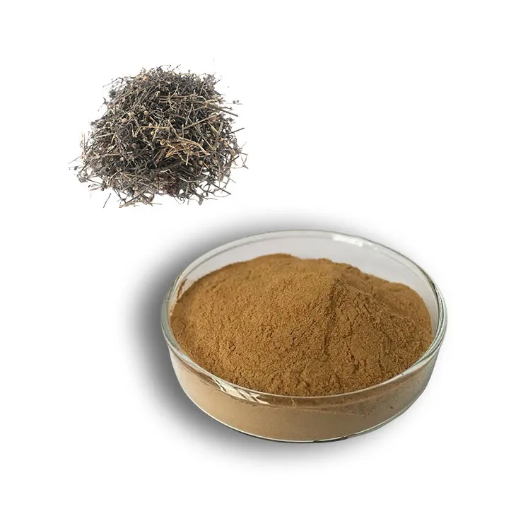
Hedyotis Diffusa Extract
2024-08-16
-
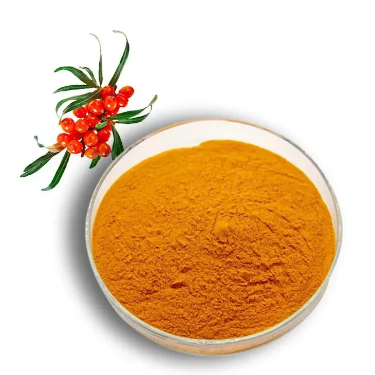
Sea buckthorn Juice Powder
2024-08-16
-
Marigold Extract
2024-08-16
-
Oat Straw Extract Powder
2024-08-16
-
Europen Bilberry Extract
2024-08-16
-
Acai Berry Extract
2024-08-16
-
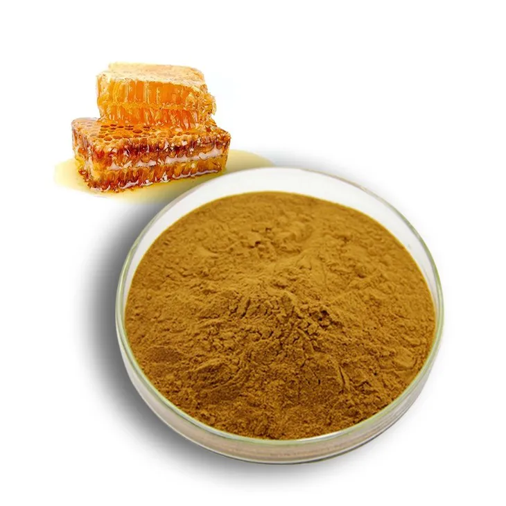
Propolis Extract Powder
2024-08-16
-
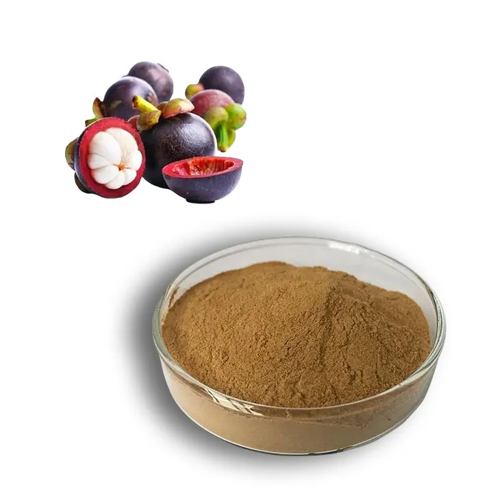
Mangosteen extract powder
2024-08-16
-
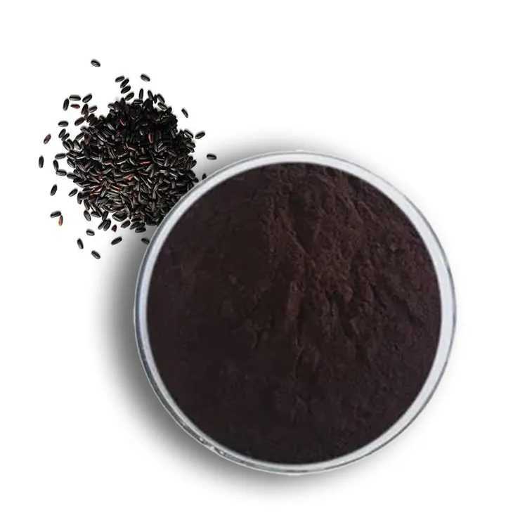
Black Rice Extract
2024-08-16
-
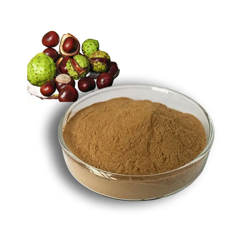
Horse Chestnut Extract
2024-08-16
