- 0086-571-85302990
- sales@greenskybio.com


Advanced Techniques in Plant Protein Extraction: Maximizing Efficiency and Yield
2024-08-03
I. Introduction
In recent years, there has been an increasing demand for plant - based proteins. This is driven by several factors, including the growing awareness of health benefits associated with plant - based diets, environmental concerns, and the need for sustainable protein sources. Plant proteins offer a viable alternative to animal - based proteins, as they are generally lower in saturated fats, cholesterol - free, and can be produced with a lower environmental footprint. As a result, the extraction of plant proteins has become a crucial area of research, aiming to meet this rising demand efficiently.
II. Mechanical Disruption Methods
A. Grinding
Grinding is one of the most common mechanical disruption methods used in plant protein extraction. It involves the use of mills or grinders to break down plant materials into smaller particles. This process helps to release the proteins trapped within the plant cells. Different types of mills can be used, such as ball mills, hammer mills, and disc mills. For example, ball mills use small balls to grind the plant material by impact and attrition. The choice of mill depends on the nature of the plant material and the desired particle size. However, grinding can generate heat, which may denature the proteins if not carefully controlled. To mitigate this, cooling systems can be incorporated during the grinding process.
B. Homogenization
Homogenization is another effective mechanical method. It uses high - pressure to disrupt plant cells. A homogenizer forces the plant material through a narrow gap at high pressure, causing the cells to rupture. This method is particularly useful for soft plant tissues. For instance, in the extraction of proteins from legumes, homogenization can effectively break down the cell walls and release the proteins. One advantage of homogenization is that it can achieve a more uniform particle size distribution compared to grinding. However, like grinding, it also requires careful control of operating parameters to prevent protein denaturation.
III. Chemical Extraction Methods
A. Solvent - based Extraction
Solvent - based extraction involves the use of solvents to dissolve and extract plant proteins. Common solvents used include ethanol, acetone, and hexane. These solvents can disrupt the lipid - protein interactions in the plant cells, thereby releasing the proteins. For example, in the extraction of oilseed proteins, hexane is often used to first remove the lipids, and then other solvents can be used to extract the proteins. However, the use of organic solvents has some drawbacks. They are often flammable, toxic, and require careful handling and disposal to avoid environmental pollution.
B. Acid - base Extraction
Acid - base extraction utilizes the solubility of proteins at different pH values. Proteins have different solubilities depending on the pH of the solution. By adjusting the pH of the extraction medium, proteins can be selectively dissolved or precipitated. For example, at a low pH (acidic conditions), some plant proteins may be soluble, while at a high pH (basic conditions), others may be soluble. After extraction, the pH can be adjusted again to precipitate the proteins. However, extreme pH values can also cause protein denaturation, so careful optimization of the pH range is necessary.
IV. Enzymatic Extraction
Enzymatic extraction is a more specific and gentle method compared to mechanical and chemical methods. Enzymes are used to break down the cell walls and membranes of plant cells, releasing the proteins without causing excessive damage to the proteins themselves.
A. Cellulase and Pectinase
Cellulase and pectinase are commonly used enzymes in plant protein extraction. Cellulase breaks down cellulose, which is a major component of plant cell walls, while pectinase hydrolyzes pectin. By using these enzymes, the cell walls can be effectively degraded, allowing the proteins to be released. For example, in the extraction of proteins from fruits and vegetables, the combined use of cellulase and pectinase can significantly improve the extraction yield. The enzymatic reaction is usually carried out under mild conditions of temperature and pH to ensure the activity of the enzymes and the integrity of the proteins.
B. Protease Inhibition
One challenge in enzymatic extraction is the presence of proteases in the plant material. Proteases can degrade the target proteins during the extraction process. To overcome this, protease inhibitors can be added. These inhibitors bind to the proteases and prevent them from acting on the proteins of interest. This helps to maintain the quality and quantity of the extracted proteins.
V. Emerging Biotechnological Approaches
With the development of biotechnology, new approaches for plant protein extraction are emerging.
A. Genetic Engineering
Genetic engineering can be used to modify plants to enhance protein extraction. For example, genes encoding for proteins with higher solubility or easier extraction can be introduced into plants. This can lead to plants that are more amenable to protein extraction processes. Additionally, genetic engineering can be used to reduce the levels of anti - nutritional factors in plants, which can interfere with protein extraction and utilization.
B. Fermentation - assisted Extraction
Fermentation - assisted extraction is another novel approach. Microorganisms can be used to ferment plant materials. During fermentation, the microorganisms produce enzymes that can break down the plant cell walls and release the proteins. Moreover, fermentation can also modify the properties of the proteins, making them more suitable for certain applications. For example, in the production of some functional food proteins, fermentation - assisted extraction can improve the bioavailability and functionality of the proteins.
VI. Process Optimization
Optimizing the extraction process is crucial for maximizing efficiency and yield.
A. Temperature
Temperature plays a significant role in plant protein extraction. Different extraction methods have different optimal temperature ranges. For mechanical disruption methods, excessive heat generation should be avoided as it can lead to protein denaturation. In enzymatic extraction, the enzymes have specific temperature optima for their activity. For example, most enzymes used in plant protein extraction have an optimal activity temperature in the range of 30 - 50 °C. Deviating from this range can reduce the enzyme activity and thus the extraction efficiency.
B. pH
As mentioned earlier, pH affects the solubility and stability of proteins. In acid - base extraction, the proper pH range needs to be determined based on the characteristics of the target proteins. In enzymatic extraction, the pH also affects the activity of the enzymes. For instance, cellulase and pectinase usually have optimal pH values in the acidic to slightly basic range. Maintaining the appropriate pH during the extraction process is essential for obtaining high - quality and high - yield proteins.
C. Extraction Time
The extraction time is another important parameter. Too short an extraction time may result in incomplete protein release, while too long a time may lead to protein degradation or other unwanted reactions. For example, in solvent - based extraction, leaving the plant material in the solvent for an extended period may cause the extraction of unwanted components or the degradation of the proteins. Therefore, the optimal extraction time needs to be determined for each extraction method and plant material.
VII. Potential Applications of Extracted Plant Proteins
The extracted plant proteins have a wide range of potential applications.
A. Food Industry
In the food industry, plant proteins can be used as ingredients in various products. They can be used to replace animal proteins in meat analogs, such as plant - based burgers and sausages. Plant proteins can also be added to dairy - like products, such as plant - based yogurts and cheeses, to improve their nutritional value. Moreover, they can be used in bakery products to enhance their texture and functionality.
B. Pharmaceutical Industry
In the pharmaceutical industry, plant proteins can be used for the development of drugs and therapeutics. Some plant proteins have biological activities that can be exploited for medicinal purposes. For example, certain plant - derived peptides have antioxidant, anti - inflammatory, or antimicrobial properties. These proteins can be purified and formulated into drugs or used as starting materials for the synthesis of new drugs.
C. Other Sectors
Plant proteins also have applications in other sectors. In the cosmetic industry, they can be used in skincare products due to their moisturizing and nourishing properties. In the biofuel industry, plant proteins can be converted into biofuels through certain biochemical processes. Additionally, they can be used in the production of biodegradable plastics, contributing to the development of sustainable materials.
VIII. Conclusion
In conclusion, advanced techniques in plant protein extraction play a vital role in meeting the increasing demand for plant - based proteins. Mechanical disruption methods, chemical extraction methods, enzymatic extraction, and emerging biotechnological approaches all offer different ways to maximize efficiency and yield. Process optimization in terms of temperature, pH, and extraction time is essential for obtaining high - quality proteins. The extracted plant proteins have diverse potential applications in food, pharmaceuticals, and other sectors. Continued research in this area is necessary to further improve the extraction techniques and expand the applications of plant proteins.
FAQ:
What are the main mechanical disruption methods in plant protein extraction?
Some of the main mechanical disruption methods in plant protein extraction include grinding, homogenization, and sonication. Grinding breaks down the plant material into smaller particles, which helps in releasing the proteins. Homogenization uses high - pressure or high - speed mixing to disrupt the cell walls and membranes. Sonication uses ultrasonic waves to create cavitation bubbles that can rupture the cells and release the proteins.
How do emerging biotechnological approaches enhance plant protein extraction?
Emerging biotechnological approaches can enhance plant protein extraction in several ways. For example, genetic engineering can be used to modify plants to produce higher levels of specific proteins or to have more easily extractable proteins. Enzyme - based methods are also emerging, where specific enzymes are used to break down cell walls more efficiently, allowing for better protein release. Additionally, some biotechnological approaches can improve the solubility and stability of the extracted proteins.
What is the significance of process optimization in plant protein extraction?
Process optimization is crucial in plant protein extraction. Parameters such as temperature, pH, and extraction time directly affect the efficiency and yield. The right temperature can influence the activity of enzymes involved in cell wall breakdown and protein solubility. The pH affects the charge and stability of the proteins. If the pH is not optimal, proteins may precipitate or become denatured. Extraction time needs to be balanced; too short may result in incomplete protein extraction, while too long may lead to protein degradation.
What are the potential applications of extracted plant proteins in the food sector?
In the food sector, extracted plant proteins can be used in a variety of ways. They can be used as meat substitutes in products like veggie burgers, sausages, and nuggets. Plant proteins can also be added to fortify foods such as bread, cereals, and dairy - like products. They can improve the nutritional profile by providing essential amino acids. Additionally, they can be used to modify the texture of food products, for example, to create a more creamy or chewy texture.
How are the extracted plant proteins used in the pharmaceutical industry?
In the pharmaceutical industry, plant - extracted proteins can be used as therapeutic agents. Some plant proteins have antibacterial, antiviral, or anti - inflammatory properties. They can be developed into drugs or used as part of drug delivery systems. Plant proteins can also be used in the production of vaccines. For example, they can be used as carriers or adjuvants to enhance the immune response. Additionally, they may be used in the development of biopharmaceuticals for treating various diseases.
Related literature
- Advanced Methods for Plant Protein Isolation and Characterization"
- "Optimizing Plant Protein Extraction: A Review of Current Techniques"
- "The Role of Biotechnology in Enhancing Plant Protein Extraction for Industrial Applications"


- ▶ Hesperidin
- ▶ citrus bioflavonoids
- ▶ plant extract
- ▶ lycopene
- ▶ Diosmin
- ▶ Grape seed extract
- ▶ Sea buckthorn Juice Powder
- ▶ Beetroot powder
- ▶ Hops Extract
- ▶ Artichoke Extract
- ▶ Reishi mushroom extract
- ▶ Astaxanthin
- ▶ Green Tea Extract
- ▶ Curcumin Extract
- ▶ Horse Chestnut Extract
- ▶ Other Problems
- ▶ Boswellia Serrata Extract
- ▶ Resveratrol Extract
- ▶ Marigold Extract
- ▶ Grape Leaf Extract
- ▶ blog3
- ▶ blog4
- ▶ blog5
-

Sugarcane Extract
2024-08-03
-

Garcinia Cambogia Extract
2024-08-03
-

Peppermint Extract Powder
2024-08-03
-
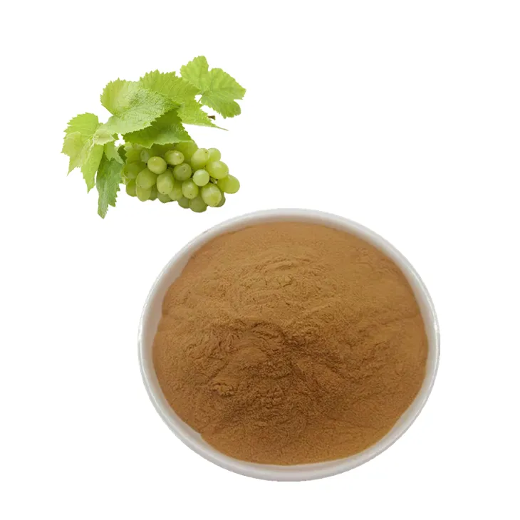
Grape Leaf Extract
2024-08-03
-
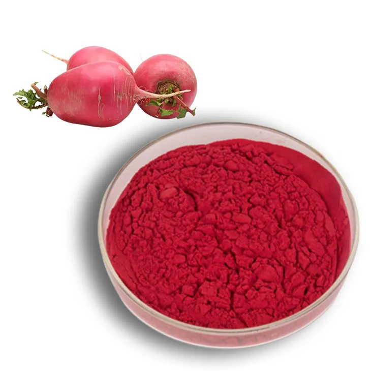
Beetroot Powder
2024-08-03
-
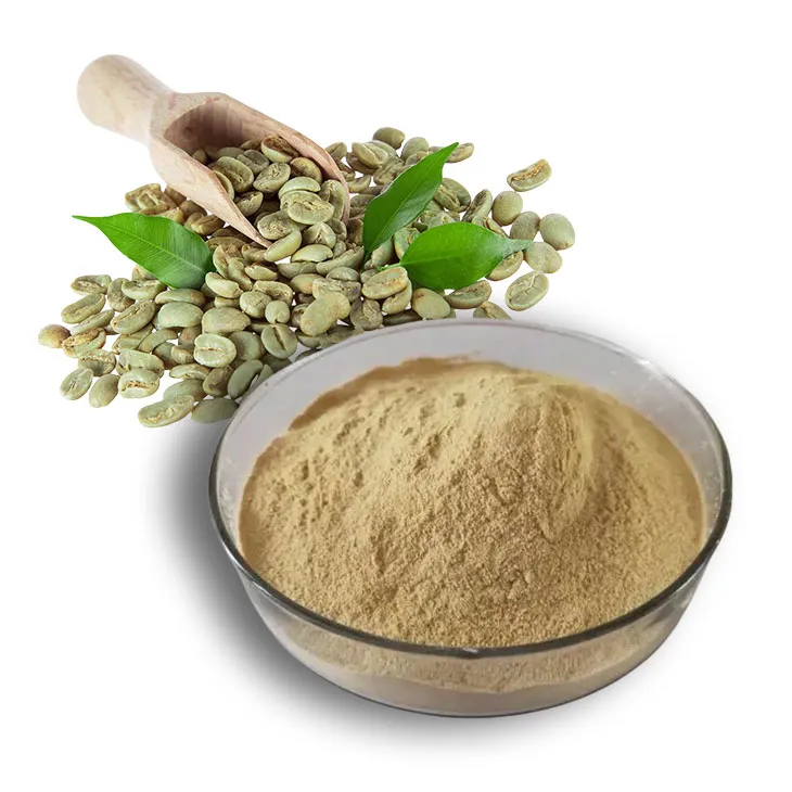
Green coffee bean Extract
2024-08-03
-
Eucommia Ulmoides Extract
2024-08-03
-
Phyllanthus Emblica Extract
2024-08-03
-
Phellodendron Extract
2024-08-03
-
White mustard seed extract
2024-08-03
