- 0086-571-85302990
- sales@greenskybio.com


From Nature to Nanotechnology: A Comprehensive Study on Plant Extract Nanoparticles
2024-08-09
1. Introduction
In recent years, the field of nanotechnology has witnessed a remarkable convergence with natural resources, particularly plant - based substances. Plant extract nanoparticles have emerged as a fascinating area of study, holding great potential for a wide range of applications. This comprehensive study aims to explore the various aspects of plant extract nanoparticles, from their extraction methods to their structural and functional features at the nanoscale, and ultimately, their implications in important areas such as drug delivery.
2. Extraction of Plant Components for Nanoparticle Synthesis
2.1 Selection of Plant Sources
The first step in obtaining plant extract nanoparticles is the careful selection of plant sources. Different plants offer a diverse range of bioactive compounds that can be utilized for nanoparticle synthesis. For example, plants like Azadirachta indica (neem) are rich in various phytochemicals. These plants are chosen based on their historical medicinal use, availability, and the known properties of their bioactive components.
2.2 Extraction Methods
There are several methods for extracting plant components for nanoparticle synthesis:
- Solvent extraction: This is one of the most common methods. A suitable solvent, such as ethanol or methanol, is used to extract the bioactive compounds from the plant material. The plant material is typically dried and ground into a fine powder before the extraction process. For instance, in the extraction of flavonoids from plants, ethanol can be an effective solvent. The solvent is then evaporated to obtain a concentrated extract containing the desired plant components.
- Supercritical fluid extraction: This method uses supercritical fluids, such as supercritical carbon dioxide. Supercritical fluids have properties between those of a liquid and a gas. They can penetrate the plant material more effectively than normal solvents and can selectively extract certain compounds. This method is often preferred for extracting heat - sensitive compounds as it operates at relatively low temperatures.
- Microwave - assisted extraction: In this method, microwave energy is used to enhance the extraction process. Microwaves can heat the plant - solvent mixture rapidly and uniformly, leading to a faster extraction time. This method is energy - efficient and can also improve the yield of the extracted compounds.
3. Structural and Functional Features of Plant - Extract Nanoparticles at the Nanoscale
3.1 Size and Shape
Plant - extract nanoparticles typically have a size range from a few nanometers to several hundred nanometers. Their size can be controlled during the synthesis process. The shape of these nanoparticles can vary widely, including spherical, rod - like, and irregular shapes. For example, silver nanoparticles synthesized using plant extracts can be spherical with an average diameter of 20 - 50 nanometers. The size and shape of the nanoparticles play a crucial role in determining their properties and applications. Smaller nanoparticles often have a larger surface - to - volume ratio, which can enhance their reactivity and interaction with other substances.
3.2 Surface Properties
The surface of plant - extract nanoparticles is rich in functional groups derived from the plant bioactive compounds. These functional groups can include hydroxyl (-OH), carboxyl (-COOH), and amino (-NH₂) groups. These surface groups can influence the stability, solubility, and biocompatibility of the nanoparticles. For example, the presence of carboxyl groups on the surface of nanoparticles can make them more negatively charged, which can affect their interaction with positively charged molecules in biological systems. Moreover, the surface of the nanoparticles can be further modified to attach specific molecules, such as drugs or targeting ligands, for various applications.
3.3 Optical and Electrical Properties
Plant - extract nanoparticles can exhibit unique optical and electrical properties. Some nanoparticles, such as gold nanoparticles synthesized from plant extracts, can show strong plasmonic resonance in the visible range of the electromagnetic spectrum. This property can be utilized for sensing applications, where changes in the plasmonic resonance can indicate the presence of a target analyte. In terms of electrical properties, certain plant - extract nanoparticles can act as semiconductors, which can be useful in electronic devices or for catalyzing electrochemical reactions.
4. Applications in Drug Delivery
4.1 Advantages over Traditional Drug Delivery Systems
Plant - extract nanoparticles offer several advantages over traditional drug delivery systems:
- Enhanced solubility: Many drugs have poor solubility in water, which can limit their bioavailability. Plant - extract nanoparticles can encapsulate these drugs and improve their solubility, allowing for better absorption in the body. For example, hydrophobic drugs can be incorporated into the hydrophobic core of nanoparticles, while the hydrophilic surface of the nanoparticles can interact with the aqueous environment in the body.
- Targeted delivery: Nanoparticles can be modified to target specific cells or tissues in the body. By attaching targeting ligands, such as antibodies or peptides, to the surface of plant - extract nanoparticles, drugs can be delivered more precisely to the site of action. This can reduce the side effects of drugs on non - target tissues.
- Controlled release: The release of drugs from plant - extract nanoparticles can be controlled. This can be achieved by adjusting the composition and structure of the nanoparticles. For instance, a drug can be encapsulated within a nanoparticle matrix that slowly degrades over time, releasing the drug in a sustained manner.
4.2 Examples of Drug - Loaded Plant - Extract Nanoparticles
There are several examples of successful drug - loaded plant - extract nanoparticles:
- Quercetin - loaded nanoparticles: Quercetin is a flavonoid with antioxidant and anti - inflammatory properties. Nanoparticles loaded with Quercetin have been synthesized using plant extracts. These nanoparticles can improve the bioavailability of quercetin and can be targeted to inflamed tissues, potentially for the treatment of inflammatory diseases.
- Paclitaxel - loaded nanoparticles: Paclitaxel is an important chemotherapy drug. Plant - extract nanoparticles loaded with paclitaxel have shown enhanced cytotoxicity towards cancer cells compared to free paclitaxel. The nanoparticles can protect the drug from premature degradation and can deliver it more effectively to cancer cells.
5. Other Potential Applications
5.1 Antimicrobial Activity
Plant - extract nanoparticles have demonstrated significant antimicrobial activity. They can inhibit the growth of bacteria, fungi, and viruses. For example, silver nanoparticles synthesized using plant extracts have been shown to have strong antibacterial properties against both Gram - positive and Gram - negative bacteria. The antimicrobial activity is thought to be due to the interaction of the nanoparticles with the microbial cell membranes, disrupting their integrity and function.
5.2 Environmental Applications
In the field of environmental science, plant - extract nanoparticles can be used for water purification. They can adsorb heavy metals and organic pollutants from water. For instance, nanoparticles synthesized from certain plants can effectively remove mercury and lead from contaminated water sources. Additionally, these nanoparticles can be used in soil remediation, helping to degrade pesticides and other organic contaminants in the soil.
6. Challenges and Future Directions
6.1 Challenges in Synthesis and Characterization
Despite the great potential of plant - extract nanoparticles, there are several challenges in their synthesis and characterization. One challenge is the reproducibility of the synthesis process. Different batches of plant materials may contain varying amounts of bioactive compounds, which can lead to differences in the properties of the synthesized nanoparticles. Another challenge is the accurate characterization of the nanoparticles. Due to their small size, it can be difficult to precisely determine their size, shape, and surface properties using traditional analytical techniques.
6.2 Toxicity and Biocompatibility Concerns
The toxicity and biocompatibility of plant - extract nanoparticles need to be thoroughly investigated. While plant - based substances are generally considered to be safe, the transformation into nanoparticles may change their properties and potential toxicity. For example, nanoparticles may be able to penetrate cells more easily than their bulk counterparts, which could lead to unexpected interactions with cellular components. Long - term studies are required to assess the safety of plant - extract nanoparticles in vivo.
6.3 Future Directions
Future research in the field of plant - extract nanoparticles should focus on several areas:
- Improving synthesis methods: Developing more reproducible and scalable synthesis methods to ensure consistent production of high - quality nanoparticles.
- Enhancing understanding of biological interactions: Further studying how plant - extract nanoparticles interact with biological systems at the molecular level to optimize their applications in drug delivery and other fields.
- Exploring new applications: Investigating new potential applications of plant - extract nanoparticles, such as in the field of regenerative medicine or energy storage.
7. Conclusion
In conclusion, plant - extract nanoparticles represent a promising area of research at the intersection of nature and nanotechnology. Their extraction methods, structural and functional features, and potential applications, especially in drug delivery, are of great interest. However, challenges such as synthesis reproducibility, toxicity, and accurate characterization need to be addressed. With further research and development, plant - extract nanoparticles have the potential to revolutionize various fields, from medicine to environmental science.
FAQ:
What are the main extraction methods for plant components in nanoparticle synthesis?
There are several common extraction methods. One is the solvent extraction method, where suitable solvents are used to dissolve and extract the active components from plants. Another is the supercritical fluid extraction, which utilizes supercritical fluids like supercritical CO₂ to extract plant substances efficiently. Maceration is also a traditional method, in which plant materials are soaked in a solvent for a certain period to obtain the desired components.
How can we analyze the structural features of plant - extract nanoparticles at the nanoscale?
Techniques such as Transmission Electron Microscopy (TEM) can be used to directly observe the morphology and size of the nanoparticles at the nanoscale. X - ray diffraction (XRD) is helpful for analyzing the crystal structure of the nanoparticles. Fourier - transform infrared spectroscopy (FTIR) can identify the functional groups present in the nanoparticles, which gives insights into their structural composition.
What makes plant - extract nanoparticles suitable for drug delivery?
Plant - extract nanoparticles often have unique properties. They can be biocompatible, which means they are less likely to cause adverse reactions in the body. Their small size allows them to easily penetrate biological membranes and reach target cells. Additionally, they can be modified to carry drugs effectively and release the drugs in a controlled manner at the target site.
Are there any challenges in the synthesis of plant - extract nanoparticles?
Yes, there are challenges. One challenge is the reproducibility of the synthesis process. Since plant materials can vary in composition depending on factors such as growth conditions and species variation, it can be difficult to achieve consistent nanoparticle synthesis. Another challenge is the purification of the nanoparticles. Removing impurities from the plant extracts during nanoparticle formation can be a complex task.
How do plant - extract nanoparticles compare to other types of nanoparticles in terms of functionality?
Compared to some synthetic nanoparticles, plant - extract nanoparticles may have the advantage of being more environmentally friendly and biocompatible. However, in terms of some specific functions such as high - precision targeting or extreme stability, some synthetic nanoparticles may outperform them. Each type has its own strengths and weaknesses, and the choice depends on the specific application requirements.
Related literature
- Plant - Based Nanoparticles for Biomedical Applications"
- "Nanoparticle Synthesis from Plant Extracts: A Green Approach"
- "The Role of Plant - Extract Nanoparticles in Drug Delivery Systems: A Review"


- ▶ Hesperidin
- ▶ citrus bioflavonoids
- ▶ plant extract
- ▶ lycopene
- ▶ Diosmin
- ▶ Grape seed extract
- ▶ Sea buckthorn Juice Powder
- ▶ Beetroot powder
- ▶ Hops Extract
- ▶ Artichoke Extract
- ▶ Reishi mushroom extract
- ▶ Astaxanthin
- ▶ Green Tea Extract
- ▶ Curcumin Extract
- ▶ Horse Chestnut Extract
- ▶ Other Problems
- ▶ Boswellia Serrata Extract
- ▶ Resveratrol Extract
- ▶ Marigold Extract
- ▶ Grape Leaf Extract
- ▶ blog3
- ▶ blog4
- ▶ blog5
-
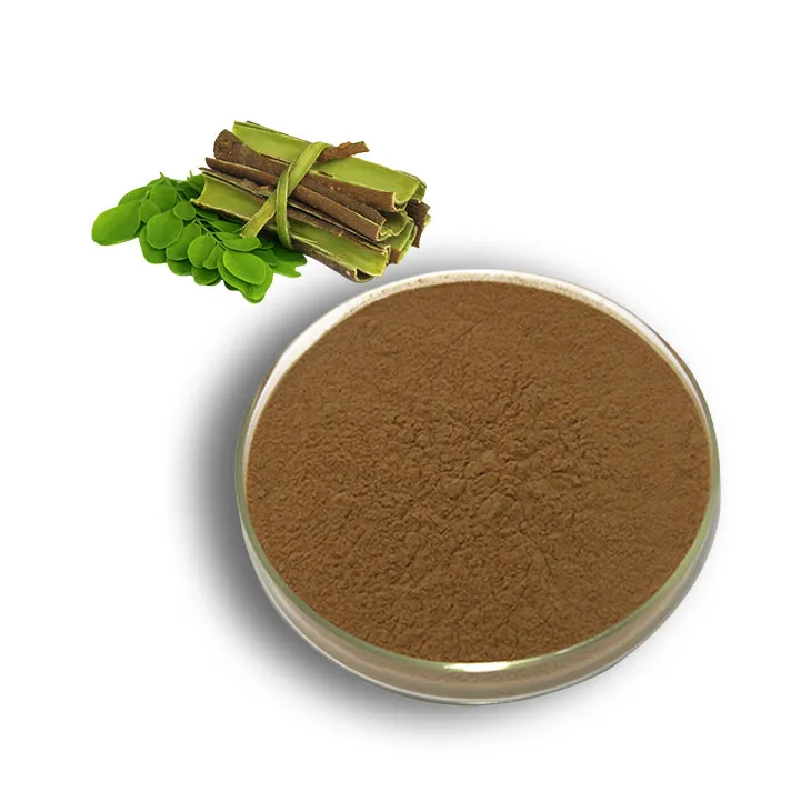
White Willow Bark Extract
2024-08-09
-
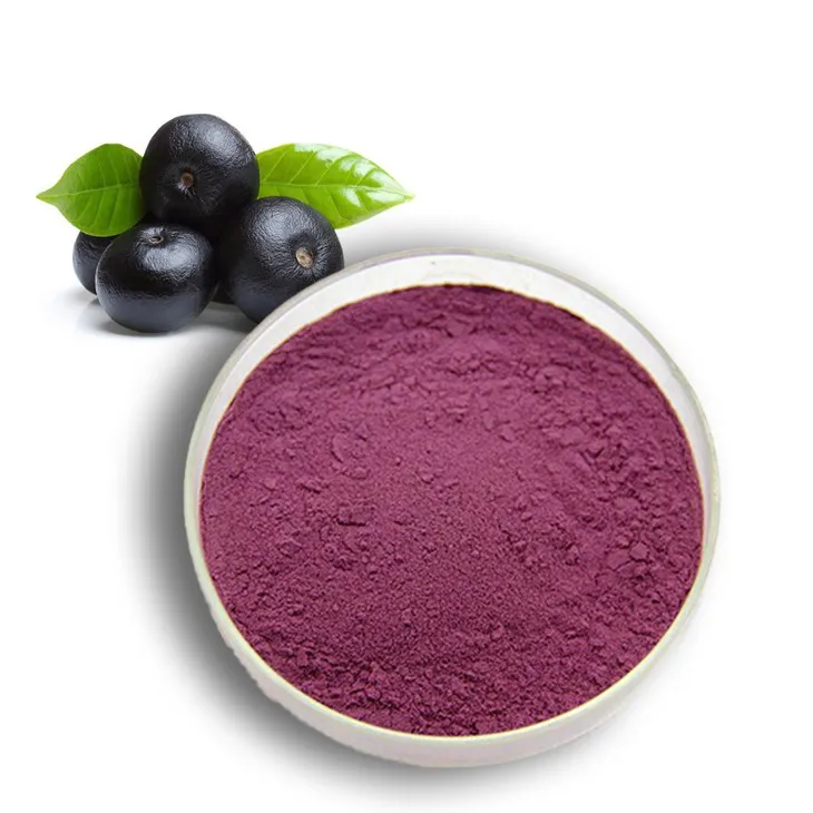
Acai Berry Extract
2024-08-09
-

Cactus Extract
2024-08-09
-
Hawthorn Extract
2024-08-09
-
Centella Asiatica Extract
2024-08-09
-
Panax Ginseng Leaf Extract
2024-08-09
-
Aguaje Extract
2024-08-09
-
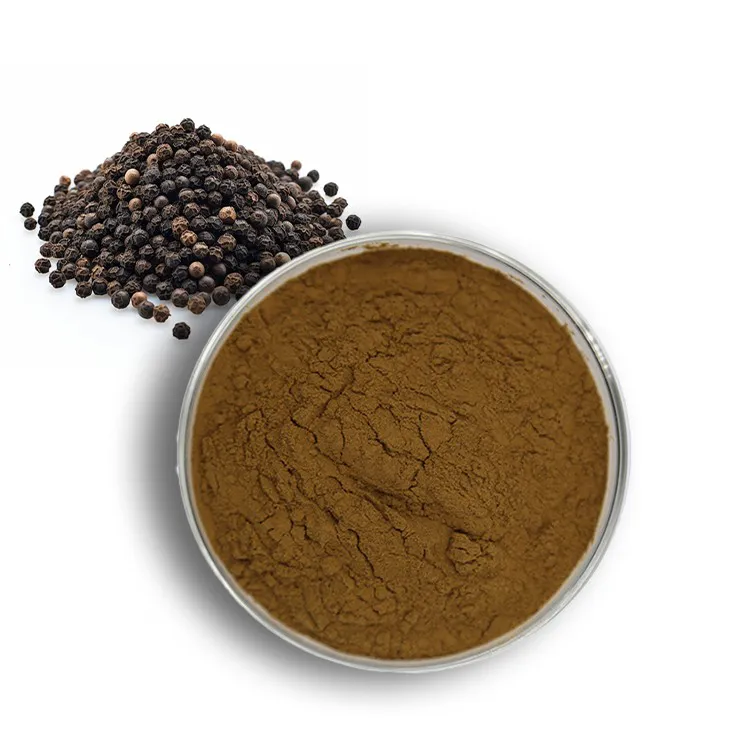
Black Pepper Extract
2024-08-09
-
Genistein
2024-08-09
-
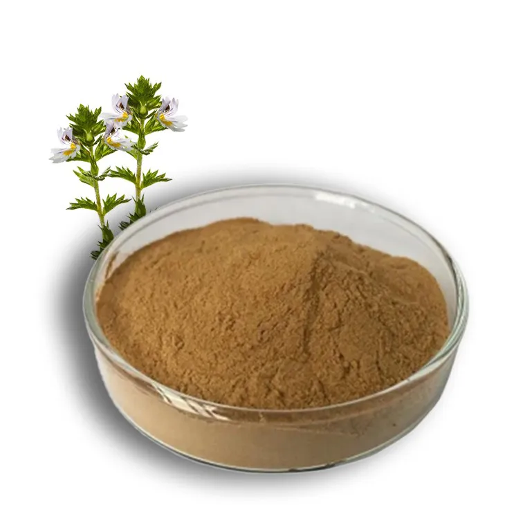
Eyebright Extract
2024-08-09
