- 0086-571-85302990
- sales@greenskybio.com


Enhancing Saponin Recovery: The Impact of Immunofluorescence on Extraction Methodologies
2024-08-06
1. Introduction
Saponins are a diverse group of natural compounds with a wide range of biological activities, including anti - inflammatory, anti - cancer, and immunomodulatory effects. Due to their potential applications in pharmaceuticals, cosmetics, and food industries, efficient extraction methods for saponins are of great importance. Immunofluorescence, a powerful technique in biological research, has recently been explored for its potential impact on Saponin Extraction methodologies. This article aims to provide a comprehensive review of how immunofluorescence can influence Saponin Extraction in various aspects.
2. Saponin Extraction: Traditional Methods
Traditional methods for saponin extraction mainly include solvent extraction, which typically uses organic solvents such as methanol, ethanol, or chloroform. These solvents are able to dissolve saponins from the plant or other natural sources. Another common method is Soxhlet extraction, which is a continuous extraction process that can provide relatively high extraction yields. However, traditional methods often have some limitations. For example, they may require large amounts of solvents, which are not only costly but also pose environmental concerns. Moreover, the extraction selectivity of traditional methods may not be optimal, leading to the co - extraction of other unwanted compounds.
3. The Basics of Immunofluorescence
Immunofluorescence is a technique that combines the specificity of antibodies with the sensitivity of fluorescence microscopy. It involves the use of fluorescently labeled antibodies to detect and localize specific antigens in cells or tissues. In the context of saponin extraction, immunofluorescence can be used to target specific saponin - related molecules or structures. The antibodies used in immunofluorescence are designed to bind specifically to the saponin or its associated components. Once bound, the fluorescent label allows for easy detection and quantification. This specificity of immunofluorescence can potentially be harnessed to improve the extraction process.
4. Impact on Extraction Yield
4.1. Selective Binding and Extraction
One of the key ways immunofluorescence can impact extraction yield is through selective binding. By using antibodies that specifically target saponins, it is possible to preferentially extract saponins from a complex mixture. For example, in a plant extract containing multiple types of secondary metabolites, the antibodies can bind only to saponins and not to other compounds such as flavonoids or alkaloids. This selective binding can lead to a higher purity of the saponin extract and potentially a higher overall yield, as less effort is wasted on extracting non - saponin components.
4.2. Optimization of Extraction Conditions
Immunofluorescence can also help in optimizing extraction conditions. Through the use of fluorescent labels, researchers can monitor the extraction process in real - time. For instance, they can observe how the saponin - antibody complexes are formed and separated during extraction. This real - time monitoring allows for the adjustment of parameters such as extraction time, temperature, and solvent composition. By optimizing these conditions based on the fluorescence signals, it is possible to maximize the extraction yield of saponins.
5. Modifying Existing Extraction Techniques
5.1. Integration with Solvent Extraction
Immunofluorescence can be integrated with traditional solvent extraction methods. In a solvent extraction process, the addition of immunofluorescence - based components can enhance the selectivity of the extraction. For example, the antibody - saponin complexes can be more easily separated from the solvent - extract mixture using techniques such as centrifugation or filtration. This integration can not only improve the quality of the saponin extract but also reduce the complexity of the subsequent purification steps.
5.2. Application in Soxhlet - like Extractions
In Soxhlet - like extraction processes, immunofluorescence can play a role in improving the efficiency. The antibodies can be immobilized in the extraction apparatus, and as the solvent continuously circulates, the saponins can be selectively bound by the antibodies. This can lead to a more targeted extraction, reducing the extraction time and potentially increasing the yield. Additionally, the immobilized antibodies can be reused, which is an advantage in terms of cost - effectiveness.
6. Comparison with Traditional Extraction Methods in Terms of Effectiveness
6.1. Purity of the Extract
When it comes to the purity of the saponin extract, immunofluorescence - based methods have the potential to outperform traditional methods. As mentioned before, the selective binding of antibodies to saponins results in a purer extract. In traditional solvent extraction, a significant amount of impurities may be co - extracted, requiring additional purification steps. Immunofluorescence - based extraction can reduce the need for such complex purification procedures, saving time and resources.
6.2. Efficiency and Yield
In terms of efficiency and yield, immunofluorescence also shows promise. While traditional methods may have relatively stable yields under certain conditions, immunofluorescence - assisted extraction can potentially increase the yield through its selective binding and optimization of extraction conditions. Moreover, the ability to monitor the extraction process in real - time can further enhance the overall efficiency, as any problems or inefficiencies can be detected and corrected promptly.
6.3. Environmental Impact
Traditional extraction methods often rely on large amounts of organic solvents, which have a negative environmental impact. Immunofluorescence - based extraction methods, on the other hand, may require less solvent due to their higher selectivity. This reduced solvent usage can contribute to a lower environmental footprint, making immunofluorescence - based extraction more sustainable in the long run.
7. Challenges and Limitations
7.1. Antibody Production and Cost
One of the major challenges in applying immunofluorescence to saponin extraction is the production of specific antibodies. Developing antibodies that can accurately and specifically bind to saponins can be a complex and time - consuming process. Additionally, the cost of antibody production can be relatively high, which may limit the large - scale application of immunofluorescence - based extraction methods.
7.2. Complexity of the Technique
Immunofluorescence is a relatively complex technique that requires specialized equipment and trained personnel. Compared to traditional extraction methods, which are often relatively straightforward in operation, the implementation of immunofluorescence - based extraction may require more investment in terms of infrastructure and training. This complexity can also lead to potential errors in the extraction process if not properly managed.
8. Future Perspectives
Despite the challenges and limitations, the potential of immunofluorescence in enhancing saponin extraction is significant. Future research could focus on developing more cost - effective antibody production methods, which would make immunofluorescence - based extraction more accessible. Additionally, efforts could be made to simplify the immunofluorescence technique for saponin extraction, reducing the complexity and the need for specialized equipment. There is also room for exploring new combinations of immunofluorescence with other emerging extraction technologies to further improve the extraction of saponins.
9. Conclusion
In conclusion, immunofluorescence has a notable impact on saponin extraction methodologies. It can influence extraction yield, modify existing extraction techniques, and offer advantages over traditional methods in terms of effectiveness. However, challenges such as antibody production cost and technique complexity need to be addressed. With further research and development, immunofluorescence - based saponin extraction has the potential to become a more widely used and efficient method in the future, contributing to the increased availability of high - quality saponin extracts for various applications in the pharmaceutical, cosmetic, and food industries.
FAQ:
Question 1: What is the significance of enhancing saponin recovery?
Enhancing saponin recovery is significant for several reasons. Saponins have various applications in the pharmaceutical, food, and cosmetic industries. Higher recovery rates can lead to more cost - effective production processes, ensure a sufficient supply for research and commercial use, and potentially unlock new applications due to increased availability of these valuable compounds.
Question 2: How does immunofluorescence influence extraction yield?
Immunofluorescence can influence extraction yield in multiple ways. It may help in specifically targeting saponin - containing structures within the source material. By binding to saponin - related antigens, it could enhance the extraction efficiency, potentially leading to a higher yield. It might also protect saponins from degradation during the extraction process, thus contributing to an increased final yield.
Question 3: Can immunofluorescence modify existing extraction techniques?
Yes, immunofluorescence has the potential to modify existing extraction techniques. It can introduce a new level of specificity to the extraction process. For example, it could be used to develop affinity - based extraction methods where the immunofluorescence - mediated binding to saponins can be exploited. This may lead to more precise and efficient extraction compared to traditional non - specific extraction techniques.
Question 4: How does the effectiveness of immunofluorescence - based extraction compare to traditional extraction methods?
The effectiveness of immunofluorescence - based extraction compared to traditional methods can vary. Immunofluorescence - based extraction offers the advantage of specificity, which may result in a purer saponin extract. However, traditional methods may be more straightforward and less expensive in some cases. In terms of yield, immunofluorescence - based extraction may potentially outperform traditional methods if properly optimized, but it also requires more complex procedures and specialized equipment.
Question 5: Are there any limitations to using immunofluorescence in saponin extraction?
There are limitations to using immunofluorescence in saponin extraction. One limitation is the cost associated with immunofluorescence reagents and the equipment required for its detection. Additionally, the development of specific antibodies or probes for immunofluorescence can be time - consuming and challenging. There may also be issues related to cross - reactivity with other compounds in the source material, which could affect the purity of the saponin extract.
Related literature
- Immunofluorescence Techniques in Natural Product Extraction"
- "Saponin Recovery: New Approaches and Traditional Methods"
- "The Role of Specificity in Saponin Extraction: Immunofluorescence Insights"


- ▶ Hesperidin
- ▶ citrus bioflavonoids
- ▶ plant extract
- ▶ lycopene
- ▶ Diosmin
- ▶ Grape seed extract
- ▶ Sea buckthorn Juice Powder
- ▶ Beetroot powder
- ▶ Hops Extract
- ▶ Artichoke Extract
- ▶ Reishi mushroom extract
- ▶ Astaxanthin
- ▶ Green Tea Extract
- ▶ Curcumin Extract
- ▶ Horse Chestnut Extract
- ▶ Other Problems
- ▶ Boswellia Serrata Extract
- ▶ Resveratrol Extract
- ▶ Marigold Extract
- ▶ Grape Leaf Extract
- ▶ blog3
-

Acai Berry Extract
2024-08-06
-

Medicinal Marshmallow Extract
2024-08-06
-
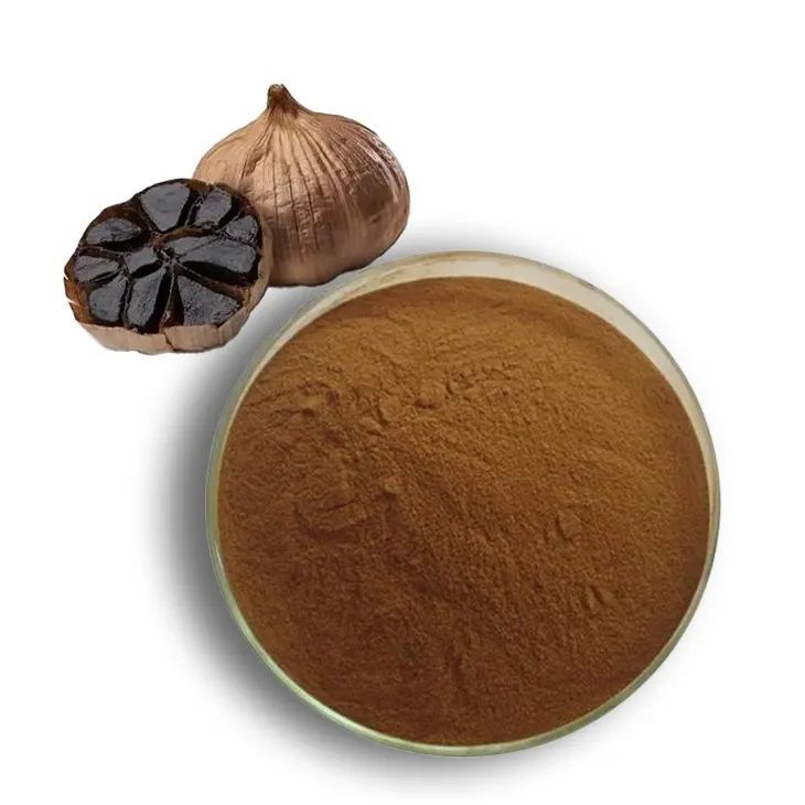
Black Garlic Extract
2024-08-06
-

Shikonin
2024-08-06
-

Nettle Root Extract
2024-08-06
-

Aminolevulinic acid
2024-08-06
-
Chaste Berry Extract
2024-08-06
-
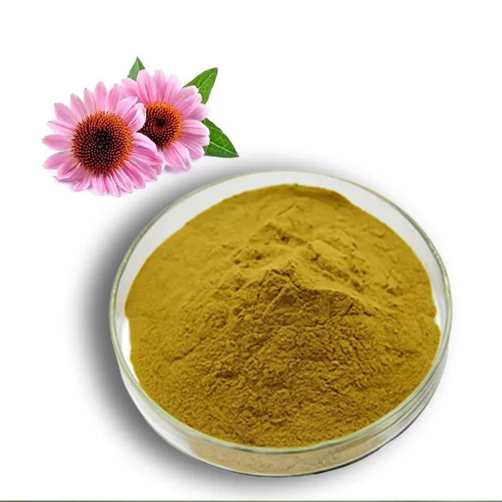
Echinacea Extract
2024-08-06
-

Aguaje Extract
2024-08-06
-
Angelica sinensis extract
2024-08-06
