- 0086-571-85302990
- sales@greenskybio.com


From Lab to Field: Streamlining Plant Tissue RNA Extraction for Robust Research
2024-08-17
1. Introduction
In the domain of plant research, RNA extraction from plant tissues is an indispensable step. RNA serves as a crucial molecule that conveys genetic information from DNA to the protein - synthesizing machinery within cells. The quality and quantity of RNA obtained play a pivotal role in a variety of downstream applications. These applications include gene expression analysis, which helps in understanding how genes are regulated under different environmental conditions or at different developmental stages of plants. Additionally, genetic engineering also heavily relies on high - quality RNA for techniques such as RNA - mediated gene silencing and gene over - expression studies.
2. Significance of Efficient RNA Extraction
2.1 Quality of RNA for Downstream Applications For gene expression analysis, accurate quantification of gene transcripts depends on the integrity of the RNA sample. High - quality RNA with intact ribosomal RNA bands and minimal degradation is essential for reliable results. In techniques like reverse - transcription polymerase chain reaction (RT - PCR), degraded RNA can lead to inaccurate amplification of gene fragments, resulting in false - negative or false - positive results. Similarly, in next - generation sequencing (NGS) technologies for transcriptome analysis, RNA quality can significantly impact the sequencing depth and coverage.
2.2 Quantity of RNA for Downstream Applications In genetic engineering experiments, sufficient RNA quantity is often required. For instance, in the case of RNA - induced gene silencing, a large amount of double - stranded RNA (dsRNA) may need to be introduced into plants. If the initial RNA extraction yields a low quantity, it may not be possible to generate enough dsRNA for effective gene silencing. Moreover, in gene over - expression studies, adequate RNA is needed for reverse transcription to complementary DNA (cDNA), which is then used for cloning and expression in transgenic plants.
3. Challenges in RNA Extraction from Plant Tissues
3.1 Presence of Secondary Metabolites Plants are rich in secondary metabolites such as polyphenols, polysaccharides, and lipids. These substances can interfere with the RNA extraction process. Polyphenols, for example, can oxidize and bind to RNA, leading to its degradation. Polysaccharides, on the other hand, can co - precipitate with RNA, reducing the purity of the RNA sample. In some plant species, high lipid content can also cause problems during extraction, as lipids can form emulsions and make it difficult to separate RNA from other cellular components.
3.2 Cell Wall Complexity Plant cells are surrounded by a rigid cell wall, which poses a significant challenge during RNA extraction. The cell wall needs to be effectively disrupted to release the intracellular RNA. Different plants have cell walls with varying compositions and thicknesses. For instance, the cell walls of woody plants are generally thicker and more lignified compared to those of herbaceous plants. This makes it more difficult to break open the cells of woody plants without causing excessive damage to the RNA.
3.3 RNA Enzyme Activity RNases are enzymes that degrade RNA. These enzymes are ubiquitous in the environment and are also present within plant cells. Once the plant tissue is disrupted during extraction, RNases can be released and start degrading the RNA. Contamination with exogenous RNases can also occur if proper precautions are not taken, such as using non - sterile equipment or reagents.
4. Optimizing RNA Extraction in the Laboratory
4.1 Choice of Extraction Method There are several methods available for RNA extraction from plant tissues, such as the phenol - chloroform extraction method, the guanidinium - based methods, and commercial RNA extraction kits. The phenol - chloroform method has been widely used for many years. It involves the use of phenol and chloroform to separate RNA from other cellular components. However, this method is time - consuming and requires careful handling of hazardous chemicals. Guanidinium - based methods, like the guanidinium thiocyanate - phenol - chloroform extraction, are more efficient in disrupting plant cells and inactivating RNases. Commercial RNA extraction kits are also popular as they are convenient and often provide high - quality RNA. These kits usually come with optimized buffers and protocols for different types of plant tissues.
4.2 Sample Preparation Proper sample preparation is crucial for successful RNA extraction. The plant tissue should be harvested at the appropriate time and stored immediately in the right conditions. For example, if the tissue is not going to be processed immediately, it should be frozen in liquid nitrogen to prevent RNA degradation. Additionally, the tissue should be ground to a fine powder in the presence of a suitable grinding agent, such as liquid nitrogen or a buffer - containing grinding buffer. This helps in efficient cell disruption and release of RNA.
4.3 RNase Inhibition To prevent RNA degradation by RNases, several measures can be taken. Firstly, all the equipment and reagents used should be RNase - free. This can be achieved by treating the equipment with RNase - inactivating agents such as DEPC (diethyl pyrocarbonate) - treated water. Secondly, RNase inhibitors can be added to the extraction buffer. These inhibitors bind to RNases and prevent them from degrading RNA. Some common RNase inhibitors include RNasin and heparin.
5. Streamlining RNA Extraction in the Field
5.1 Portable and Field - Friendly Extraction Kits In field settings, it is often necessary to extract RNA on - site. Portable RNA extraction kits have been developed to meet this need. These kits are small in size, easy to carry, and can be used with minimal equipment. They are designed to work under less - than - ideal conditions, such as in the presence of contaminants and without access to a full - fledged laboratory. Some field - friendly kits use dry reagents, which have a longer shelf - life and are more stable during transportation.
5.2 Field Sampling and Preservation Field sampling for RNA extraction requires careful consideration. The samples should be collected in a way that minimizes damage and RNA degradation. Special sampling tools can be used to ensure quick and clean collection of plant tissues. After sampling, the tissues need to be preserved immediately. One common method is to use RNA later, a reagent that can be added to the sample in the field to stabilize RNA. Another option is to freeze the samples using dry ice or portable freezers if available.
5.3 Minimizing Contamination in the Field In the field, there is a higher risk of contamination compared to the laboratory. To minimize contamination, clean sampling techniques should be employed. For example, wearing gloves and using sterile sampling tools can reduce the introduction of exogenous contaminants. Additionally, the sampling area should be clean and free from sources of RNase contamination, such as soil or plant debris that may contain high levels of RNases.
6. Innovative Solutions to Overcome Extraction Challenges
6.1 New Chemical Reagents Researchers have developed new chemical reagents to address the problems associated with secondary metabolites and RNA extraction. For example, some reagents are designed to specifically bind to polyphenols and remove them from the extraction mixture, thereby improving the purity of the RNA. Others are formulated to prevent polysaccharide co - precipitation with RNA. These new reagents offer more targeted solutions compared to traditional extraction methods.
6.2 Advanced Cell Disruption Techniques In addition to traditional grinding methods, new cell disruption techniques have been developed. For example, ultrasonic - assisted cell disruption can be used to break open plant cells more efficiently. This technique uses high - frequency sound waves to create cavitation bubbles within the sample, which then collapse and cause mechanical disruption of the cell walls. Another technique is the use of bead - beater, where small beads are shaken vigorously with the plant tissue to disrupt the cells. These advanced techniques can be particularly useful for plants with tough cell walls.
6.3 Integrated RNA Extraction Systems Integrated RNA extraction systems have been developed that combine multiple steps of the extraction process into a single device or protocol. These systems can streamline the extraction process, reduce the time required, and improve the reproducibility of the results. For example, some systems integrate cell disruption, RNA purification, and RNase inactivation into one continuous process.
7. Conclusion
In conclusion, efficient RNA extraction from plant tissues is of utmost importance for robust plant research. By understanding the significance of high - quality and sufficient - quantity RNA, as well as the challenges associated with the extraction process, researchers can optimize the extraction methods both in the laboratory and in the field. The development of innovative solutions, such as new chemical reagents, advanced cell disruption techniques, and integrated extraction systems, further enhances the reliability and efficiency of RNA extraction. This, in turn, will facilitate more accurate gene expression analysis, successful genetic engineering, and overall more impactful plant - related research.
FAQ:
What are the main challenges in plant tissue RNA extraction?
There are several main challenges. Firstly, plant tissues often contain high levels of polysaccharides, polyphenols and secondary metabolites which can interfere with RNA extraction. These substances can co - precipitate with RNA or cause degradation. Secondly, different plant tissues have different cell structures and compositions, which may require different extraction protocols. For example, leaf tissues may be easier to handle compared to root tissues which are rich in lignin and other complex substances. Thirdly, in field - collected samples, there may be contaminants such as soil particles and microbes which can affect the purity of the RNA extraction.
Why is efficient RNA extraction important for gene expression analysis?
Efficient RNA extraction is crucial for gene expression analysis. High - quality RNA is required for accurate reverse transcription into cDNA. If the RNA is degraded or impure, it will lead to inaccurate quantification of gene expression levels. The quantity of RNA also matters, as insufficient RNA may not be enough for certain sensitive detection methods. Moreover, pure RNA without contaminants is necessary to avoid false - positive or false - negative results in gene expression assays.
How can the RNA extraction process be optimized in field settings?
In field settings, the following steps can be used to optimize RNA extraction. First, proper sample collection and preservation are essential. Samples should be collected quickly and stored in appropriate buffers or at low temperatures to prevent RNA degradation. Second, portable and easy - to - use extraction kits can be employed. These kits are often designed to be more robust against field - related contaminants. Third, pre - treatment methods can be applied in the field to remove some of the major contaminants, such as a quick washing step to remove soil particles from plant tissues.
What are the innovative solutions to overcome challenges in plant tissue RNA extraction?
One innovative solution is the use of modified extraction buffers. These buffers are formulated to specifically deal with the interference of polysaccharides and polyphenols. For example, some buffers contain reagents that can selectively precipitate these substances away from RNA. Another solution is the development of new extraction techniques, such as magnetic - bead - based RNA extraction. This method can provide more specific and efficient capture of RNA molecules compared to traditional methods. Additionally, the use of molecular biology - grade enzymes that are more resistant to inhibitors present in plant tissues can also improve the extraction process.
How does high - quality RNA extraction contribute to genetic engineering in plants?
High - quality RNA extraction is fundamental for genetic engineering in plants. In genetic engineering, gene expression needs to be accurately controlled. RNA is used to study gene function and regulation before any genetic modification. If the RNA extraction is of poor quality, it will lead to incorrect understanding of gene function, which in turn can result in unsuccessful genetic engineering attempts. Also, during the process of creating transgenic plants, accurate RNA analysis is required to monitor the expression of introduced genes, and high - quality RNA extraction ensures reliable results in these analyses.
Related literature
- Title: Improved RNA Extraction from Plant Tissues for Advanced Genomic Studies"
- Title: "RNA Extraction Protocols for Diverse Plant Tissues: A Comprehensive Review"
- Title: "Optimizing Field - Based RNA Extraction from Plant Samples for Molecular Research"


- ▶ Hesperidin
- ▶ citrus bioflavonoids
- ▶ plant extract
- ▶ lycopene
- ▶ Diosmin
- ▶ Grape seed extract
- ▶ Sea buckthorn Juice Powder
- ▶ Beetroot powder
- ▶ Hops Extract
- ▶ Artichoke Extract
- ▶ Reishi mushroom extract
- ▶ Astaxanthin
- ▶ Green Tea Extract
- ▶ Curcumin Extract
- ▶ Horse Chestnut Extract
- ▶ Other Problems
- ▶ Boswellia Serrata Extract
- ▶ Resveratrol Extract
- ▶ Marigold Extract
- ▶ Grape Leaf Extract
- ▶ blog3
- ▶ blog4
- ▶ blog5
-
Chia Seed Powder
2024-08-17
-
Sugarcane Extract
2024-08-17
-
Chaste Berry Extract
2024-08-17
-
Hedyotis Diffusa Extract
2024-08-17
-
Aguaje Extract
2024-08-17
-
Black Garlic Extract
2024-08-17
-
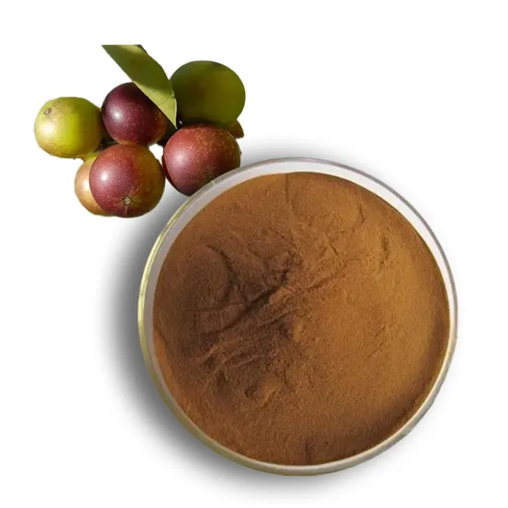
Camu Camu Extract
2024-08-17
-
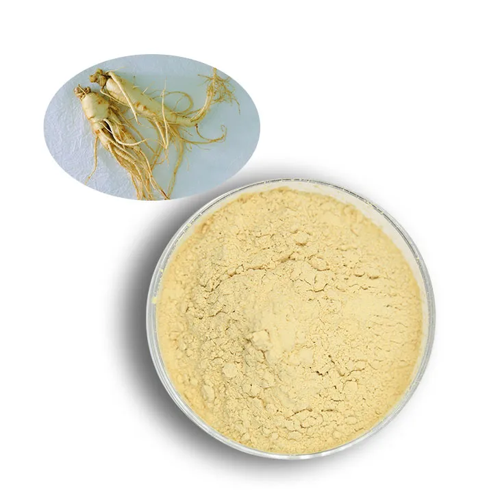
American Ginseng Root Extract
2024-08-17
-
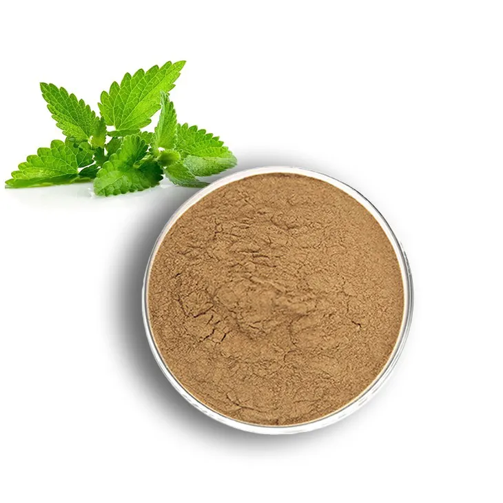
Lemon Balm Extract
2024-08-17
-
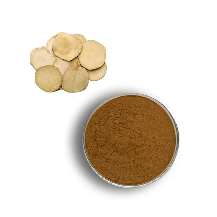
Alisma Extract
2024-08-17
